During the life cycle of a cell, each cell divides to form two genetically identical cells. This process is called mitosis. Just before the cell enters mitosis, the DNA duplicates. During the process of mitosis each half of the DNA is pulled to towards the opposite poles of the cell. When the cell divides into two, each new daughter cells receives one copy of the full DNA complement, identical to the parent.
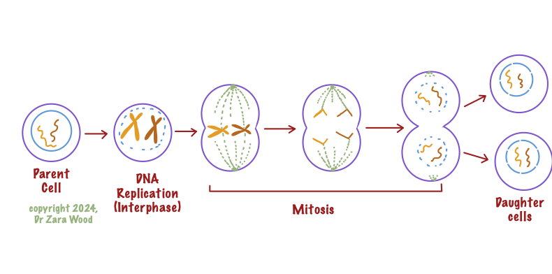
DNA is replicated in interphase, and the sister chromatids partitioned into daughter cells during Mitosis, to produce genetically-identical cells. Diploid parents give rise to diploid daughters.
Spindle fibres are key to the process of mitosis. During the process of DNA separation, spindle fibres join each half of the DNA (called a sister chromatid) to one pole of the cell.

The centrosome forms the microtubule organisation centre (MTOC). Each centrosome contains a pair of centrioles. Spindle fibres from the centrosome join the kinetochore on either side of the chromosome. The sister chromatids are aligned on the equator, one of top of the other.
The sister chromatids are joined together at the centromere. Spindle fibres are connected to the the sister chromatids at the kinetochore. At the other end spindle fibres connect to the centrioles located at the poles of the cell.
When the spindle fibres contract, each sister chromatid is pulled towards the opposite pole of the cell.
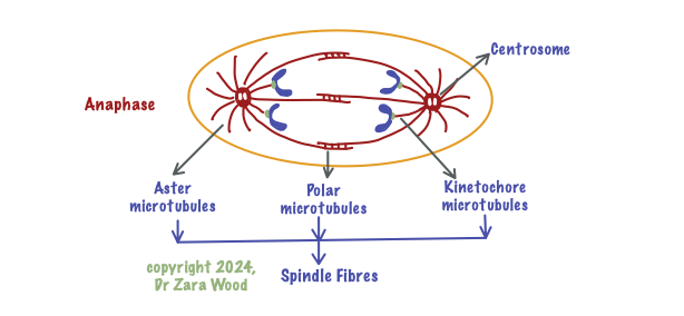
Spindle fibres are made up of three types of microtubules – aster, polar and kinetochore tubules. During Anaphase, the kinetochore microtubules contract, causing the sister chromatids to be pulled apart, to opposite poles of the cell.
Spindle fibres are made of a protein called tubulin (of which there are 2 types – alpha- and beta-tubulin). Tubulins can be added or removed from the spindle fibres. Addition of tubulin proteins causes the spindle fibres to elongate (polymerisation), and removal of the proteins causes the spindle fibres to shorten (depolymerisation).
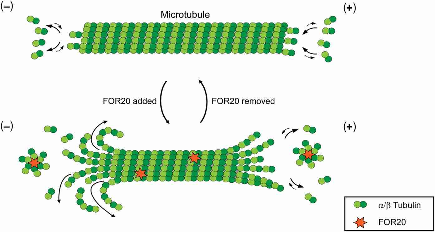
From: Microtubule-binding protein FOR20 promotes microtubule depolymerization and cell migration Feng, S., Song, Y., Shen, M. et al., Cell Discov 3, 17032 (2017). https://doi.org/10.1038/celldisc.2017.32
During spindle fibre contraction, the process of depolymerisation causes spindle fibres to shorten. Several hypotheses have been proposed to explain the movement of the chromosomes along the spindle fibres. The first is that an ATP-dependent protein, kinesin, uses the energy from ATP hydrolysis to pull the sister chromatids towards the opposite poles of the cell. The second hypothesis suggests that the energy from hydrolysing a molecule of GTP is stored in the tubulin proteins when they are being assembled, and released when they depolymerise, providing the energy for chromosome movement.

This is a real-time video of cell division, showing the spindle fibres pulling the chromosomes to the opposite poles of the cell:
